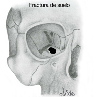
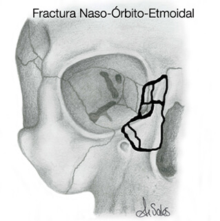
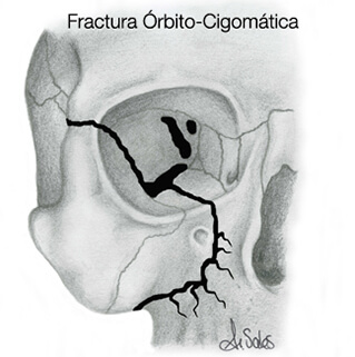
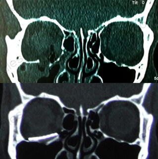
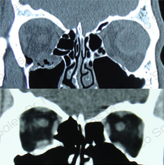
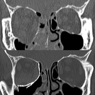
The orbit is the cavity of the face that houses the eye, the fat and the muscles that move it. It is formed by the union of different bones, and is shaped like an inverted pyramid, with the base facing outwards. Four walls can be distinguished in the orbit: the floor, the roof, the medial wall (the one facing the nose) and the lateral wall (the one facing outwards).
In orbital fractures, one or more walls may be broken (burst fractures, the most common) or the orbital rim may also be broken. Fractures involving the orbital rim usually involve much greater trauma and are less common.
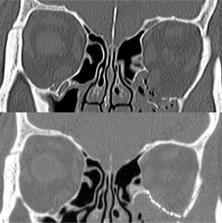
Blow-out wall fractures are typically caused by blunt blows to the eye, such as a ball. They cause the pressure inside the orbit to increase greatly and it breaks through the floor or the medial wall, which are the thinnest and least resistant walls. When the floor breaks, for example, what happens is that the orbital space or volume (“the room where the eye is”) becomes larger, as the orbit communicates with the maxillary sinus (it would be as if a partition is removed in one room and communicates with the one next door, making a much larger room).
As the space becomes larger, the fat has more room to spread out, and the eye sinks inwards, producing Enophthalmos. In addition, the inferior rectus muscle is located very close to the ground, so that when it breaks, the muscle can become trapped in the bone fragments and prevent the eye from moving normally, causing double vision or diplopia.
If a fracture does not cause double vision (diplopia) or sinking of the eye (enophthalmos), surgery is generally not necessary. If the double vision does not improve in the first few weeks or if there is enophthalmos, surgery is necessary. The surgery consists of accessing the floor of the orbit, removing the herniated tissues in the maxillary sinus or trapped in the fracture, and reconstructing the defect left behind so that the orbital tissue cannot be pushed back into the sinus. To access the floor of the orbit, the most recommended incision is the transconjunctival incision, inside the eyelid, which leaves no visible scar and is also the least likely to cause retraction of the lower eyelid (leaving the lower eyelid displaced downwards, making it more difficult to close the eyes). The material that can be used to reconstruct the floor of the orbit can be the patient’s own bone or titanium mesh.

